 |
|||||||||||||||||||||||||||||||||||||||||||||||
|
|
|||||||||||||||||||||||||||||||||||||||||||||||
       |
Special Feature: Nanobiotechnology Research Vol. 7, No. 8, pp. 5–9, Aug. 2009. https://doi.org/10.53829/ntr200908sf1 Nanobiotechnology Research at NTTAbstractThis article introduces nanobiotechnology research being conducted at NTT as an introduction to the Special Feature in this issue. The other articles describe individual topics in more detail. One of the principal mechanisms of memory and learning is considered to be synaptic plasticity. We are interested in understanding this mechanism and using it to develop a novel information processing system that will be useful for telecommunication networks of the future. 
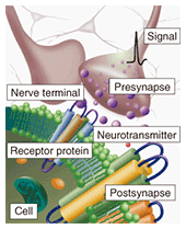
1. IntroductionA synapse is a nerve cell terminal that transmits electrical signals to the corresponding neuron (Fig. 1). At this terminal, an electrical signal is converted into a chemical signal, such as a neurotransmitter. Information is coded by changing either the release amount or release frequency of the neurotransmitter and modified by changing the receptor sensitivity at the synapse. The main neurotransmitters in the brain are glutamate and gamma-aminobutyric acid (GABA). They are released from presynaptic terminals into a region about 100 nm in size known as the synaptic cleft, and they move towards postsynaptic cells, which contain receptors that receive the neurotransmitters as information. There has been considerable discussion about whether neurotransmitter amount/frequency or receptor sensitivity is the dominant process in synaptic plasticity, memory, and learning. Although no incontrovertible proof has yet been provided, recent results indicate that receptor sensitivity could be the key to the process.
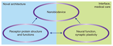
Our aim is to develop devices that can communicate with the brain using receptor proteins with nanoelectrodes and biological energy sources. The key idea behind achieving these devices is the fusion of receptor conformation/function and synaptic connection/function (Fig. 2). Such devices are regarded as nano-eyes, nano-noses, and nano-ears, according to the protein used in the device.
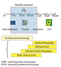
2. Nanobiotechnology/bionanotechnologyNanobiotechnology/bionanotechnology is a multidisciplinary field combining nanotechnology and biotechnology [1]–[3]. Although nanobiotechnology has been defined as relating to an understanding of biological functions and mechanisms using nanotechnology and bionanotechnology is defined as developing nanotechnology using biological phenomena and functions, these terms are now largely interchangeable. Nanobiotechnology is concerned with a wide range of research targets from molecules to bacteria and covers the nanometer-to-micrometer range. The corresponding technologies used in this research field are optical and electron beam lithography and scanning probe microscopy (Fig. 3). A typical image of this technology can be drawn from Isaac Asimov’s famous novel “Fantastic Voyage”. Although forty years have passed since its publication, we have yet to miniaturize people for a voyage inside a human body. However, we have managed to miniaturize problem-solving machines (micromachines) that can be introduced into a human body. Furthermore, drug delivery systems designed to control the molecules, medicine, and gene therapy used for treating patients could bring the achievement closer. This field also includes protein nanomotors, such as myosin and dynein, and tissue engineering such as cell sheet technology.
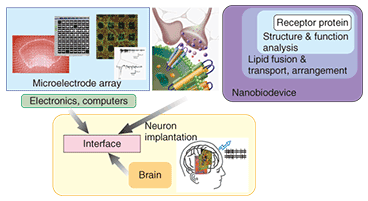
3. Research at NTTThe principal role of neural information processing in the brain can be discussed by using the following three approaches (Fig. 4): analysis of the structures of receptor proteins depending on their function (top right in Fig. 4), development of a neuron-machine interface using a microelectrode array with conductive polymer hydrogel (top left in Fig. 4), and formation of artificial synapses using the results of the first two studies [4]–[7]. These are the goals of our research. For the Special Feature in this issue, I have selected six articles that introduce our research activities.
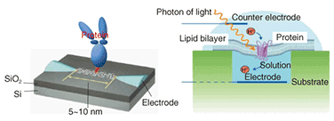
First, let me explain the concept of receptor protein visualization under physiological conditions. We are studying the conformational changes induced by neurotransmitter reception to enable us to understand the relationship between conformational change and function [8], [9]. This is very important for the development of nanobiodevices using receptor proteins. However, it is extremely difficult to visualize the conformational changes in a receptor under physiological conditions. Only a few technologies, such as X-ray diffraction and cryogenic transmission electron microscopy (CRYO-TEM) can provide us with images of receptors. This is because receptors are very small (around 10 nm) and function only in liquid. Therefore, measurements need to be performed in liquid, whereas most methods require dry conditions or low temperature. We have succeeded in visualizing these receptors under physiological conditions in real time by using high-speed atomic force microscopy. The visualization of receptor proteins and conformational analysis is explained in the article entitled “Direct Observation of Dynamics in Receptor Protein by Atomic Force Microscopy” [10]. As the receptor normally works with a membrane, the reconstitution of proteins into a membrane is a key to understanding the function under physiological conditions. This is described in “Reconstitution of Membrane Proteins into Lipid Bilayer and Its Application to Nanobiodevices” [11]. To develop nanobiodevices or bionanodevices, it is necessary to control receptor protein placement and alignment under controlled conditions. This is very difficult, but important if we are to develop a nanobiodevice architecture using receptor proteins. We are studying protein placement using either a gold nanodot array [12] or self-spreading lipid bilayers, which are regarded as artificial biological membranes because of their characteristics. The latter approach is presented in “Artificial Cell Membrane on Patterned Surface—Growth Control and Microchannel Device Application” [13]. We are also investigating the bio-sensing capability of lipid-membrane-coated gold nanorods, which have characteristic optical properties that originate from localized surface plasmons. Self-assembly means here that we can achieve various alignments in one, two, and three dimensions. This study is introduced in “Control of Gold Nanorod Arrays Through Self-assembly of Biomolecules” [14]. Research based on a nanoelectrode device that uses a conductive polymer as a molecular wire is introduced in “Nanometer-scale Analysis of Single Molecule” [15]. The fusion of receptor proteins with molecular wire is one of our targets for a receptor-based nanobiodevice architecture. Our approach to constructing an interface between neurons and electrical instruments through electrodes, thus achieving a longer connection, is described in “Neural Function Observation with Microelectrode Array” [16]. We use conducting polymer hydrogels to achieve better biocompatibility and low impedance [17]. One major challenge is to miniaturize our measurement system into an LSI (large-scale integrated circuit) chip with the aim of making chips that can be implanted into the human body in the future. Using this research as a basis, we plan to develop an artificial synapse that allows us to access our information processing systems, namely our brains. We think that strategic collaboration is one of the fastest and most promising ways to accomplish this goal. We began a collaboration with the University of Oxford in October 2004 and established a laboratory there. Some successful results are described in “Reconstitution of Membrane Proteins into Lipid Bilayer and Its Application to Nanobiodevices” [11]. We have developed a high-density self-assembled vesicle array for high-throughput protein analysis using a gold-nanodot array. This collaboration will both help us and promote our research activities through several funding schemes supported by governmental funding agencies, such as BBSRC [18], EPSRC [19], and JST [20]. These agencies are already supporting us by securing our research resources. 4. Concluding remarksRecent developments in biological research are totally dependent on developments in nanotechnology [1]–[3]. The visualization of nanostructures and certain phenomena has led to great progress in understanding protein structures and functions, and molecular interactions, which are very difficult to observe and analyze. This will help us to clarify complex phenomena, such as protein-protein interactions. The goals of our research are to understand the mechanisms of informational signal transduction, such as memory and learning, and to develop biomimetic self-standing devices using biological energy sources. We have successfully visualized receptor proteins and their conformational changes under physiological conditions. Further studies are required if we are to understand the relationship between structure and function, such as synaptic plasticity. The combination of synaptic plasticity with nanoelectrodes and reconstituted receptor proteins will provide us with bio-friendly nanobiodevices (Fig. 5) that could make it possible to clarify the neural information processing system in the brain [4], [5], [21].
In future, artificial synapses could be developed for medical applications in functional support devices and for biological signal transduction interfaces, in addition to telecommunication. Our dream is to understand the neural information processing system and to help find cures for spinal cord injuries and diseases like Alzheimer’s. References
|
||||||||||||||||||||||||||||||||||||||||||||||