 |
|||||||
|
|
|||||||
       |
Feature Articles: Healthcare Devices and Infrastructure-maintenance Technology that Support People and Society Vol. 19, No. 6, pp. 55–60, June 2021. https://doi.org/10.53829/ntr202106fa8 Non-invasive Biological-information Sensing Using Photoacoustic Measurement TechnologyAbstractBiological-information sensing technology for simplifying the measurement of blood components without drawing blood has been attracting attention. NTT has been researching and developing biological-information sensing using photoacoustic measurement technology that combines the ability of light to selectively measure specific biological components and the ability of sound (ultrasound) to propagate through the human body. In this article, we introduce non-invasive biological-information sensing technology using photoacoustic measurement technology with the aim of collecting various types of information from within the body. Keywords: photoacoustic, non-invasive, sensor
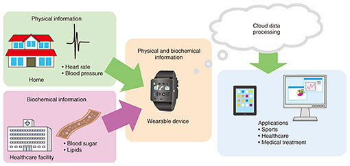
1. Advances in biological-information sensing through non-invasive measurement technologiesRecent progress in information and communications technology (ICT) and growing awareness of health management has made personalized healthcare services that use biological information obtained from wearable devices increasingly popular. Information that is monitored during an individual’s daily life shows promise for application to systems that promote behavior modification or use healthcare-oriented big data (Fig. 1). However, biological information obtained from current wearable devices consists mostly of data such as amount of activity, heart rate, respiration, body temperature, and blood pressure obtained from physical sensors. Consequently, when one is not feeling well, for example, such biological information cannot be used to describe the cause or suggest a countermeasure even if one’s state can be objectively presented. It would therefore be desirable if wearable devices could also provide biochemical information such as data on blood components. The current method of obtaining such information is to draw blood at a hospital or medical clinic and analyze that sample using various types of test equipment on an item-by-item basis. Alternatively, to obtain blood-component information from wearable devices, it would be necessary to establish technology that could easily obtain data in a continuous and non-invasive manner without harming the body. In this regard, biological imaging technology using the photoacoustic effect has recently been investigated as a non-invasive biological sensing technique. This is a powerful technique for learning about the body’s interior by combining two key features: the ability of light to measure biological components selectively and the ability of sound to propagate well through the body. In other words, the strongpoint of photoacoustic measurement technology is the ability to make measurements deep into the body that light cannot easily penetrate. There is a great need to look inside the human body at healthcare facilities, and this is currently done through the use of diagnostic imaging equipment such as X-ray computed tomography, magnetic resonance imaging, and positron emission tomography. In contrast to such diagnostic imaging equipment, photoacoustic equipment consisting of a light source and microphone has the advantage of being simple and portable with little burden placed on the user. We have been researching and developing the application of the photoacoustic effect to biochemical information measurements from the viewpoint of establishing technology that enables measurements to be conducted anytime and anywhere. In this article, we first provide a basic description of the principle behind the photoacoustic effect then introduce the current state of photoacoustic research at NTT.
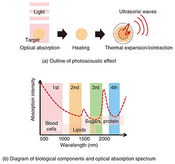
2. Measurements using the photoacoustic effectThe photoacoustic effect was discovered in 1880 by Alexander Graham Bell, the inventor of the telephone, as a phenomenon in which the intermittent irradiation of a substance with light generates sound. In 1965, Mark Leonidovitch Veingerov proposed the application of the photoacoustic effect to the analysis of gaseous components at concentrations on the ppm (parts per million) level (one-ten-thousandth of 1%), since then, techniques have been established for using the photoacoustic effect to analyze chemical structures on a molecular level. We describe the photoacoustic effect in easy-to-understand terms. As shown in Fig. 2(a), the absorption of light by matter is followed by the conversion of that optical energy to heat and the thermal expansion of that substance due to this thermal energy. If such optical irradiation should be applied in an intermittent manner, thermal expansion will occur repeatedly giving rise to elastic waves, that is, sound (ultrasonic waves). These are called photoacoustic waves. Recent advances in ICT have also led to amazing benefits from a variety of devices and technologies such as light sources and optical switches that can generate optical pulses with high temporal controllability, acoustic technologies that can detect sound with high sensitivity, and electronics technologies that can perform diverse signal processing. Such devices and technologies have been applied in a wide range of fields including material analysis, gas analysis, and imaging. In biological measurements using the photoacoustic effect, the amount of light absorbed by the target substance within the body is converted to ultrasonic waves that propagate well through the body, and the concentration of that substance is estimated by detecting those waves as photoacoustic waves. Substances have optical colors (wavelengths) that are easy to absorb due to individual characteristics. This feature can be used to learn about biological components and their distribution in the human body. For example, as shown in Fig. 2(b), blood cells, lipids, and glucose/proteins absorb light well in the 500-nm (visible light), 1000-nm (near-infrared light), and 1500-nm (near-infrared light) regions, respectively. Making use of this property, information on biological components can be obtained by irradiating the body with various colors and measuring how much of that light is absorbed. It is well known that more than half of a living body is composed of water, which also has the property of absorbing light. Consequently, when using light to conduct measurements, it is generally necessary to select optical wavelengths that are not greatly absorbed by water. This is because no wavelength absorption by water makes it easier for light to enter the body without attenuation. This range of optical wavelengths is called the “optical window.” At the same time, there are limits to the depth at which light can penetrate the body for conducting measurements, and it is extremely difficult to determine by what path and in what way light is absorbed as it passes through our bodies intricately composed of diverse components. Despite this problem, photoacoustic measurements are conducted to detect the amount of absorbed light by converting it to ultrasonic waves that propagate well through the body, which means that the effects of reflection in the body can be suppressed. It is also possible to investigate the interior of the body in a non-invasive manner by measuring not only the intensity of photoacoustic waves (sound pressure) from light absorption by the target substance but also their propagation time and frequency characteristics.
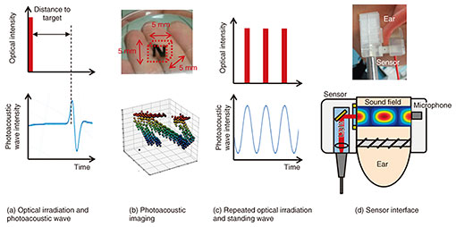
3. Non-invasive approaches for measuring biological components using the photoacoustic effectThere are two approaches for measuring biological components in a non-invasive manner using the photoacoustic effect. One involves irradiating the body with a light pulse of extremely short duration (pulse width: several nanoseconds), having the target substance absorb that light, and measuring both the time taken for the generated photoacoustic wave to return and the position of the component absorbing the light on the basis of the sound pressure of that wave. With this approach, a photoacoustic wave is obtained as shown in Fig. 3(a), and the depth of the target substance is determined from the time spent by the sound propagating through the body and its speed. A demonstration of three-dimensional imaging of the letter “N” embedded in material simulating the human body is shown in Fig. 3(b). The second approach, as shown in Fig. 3(c), involves irradiating the body with light that is periodically turned ON/OFF, letting the photoacoustic wave generated from the target substance reflect any number of times to generate a standing wave, and measuring the concentration of the target from the sound pressure of that wave. The former approach can be used to obtain information on a substance and its position. For example, knowing that many fine blood vessels form near cancer cells that require a considerable amount of nutrition to survive, much applied research has been conducted on imaging that situation. Such research, however, generally requires equipment that is large and expensive. Another problem is that signals that include information on biological components are weak and easily affected by noise. The latter approach, while not able to obtain positional information of the target substance, can obtain a relatively large signal-to-noise ratio with just a low-intensity, inexpensive light source and one microphone, which makes it advantageous for measuring component concentrations or downsizing equipment. However, component measurements cannot provide clear contours such as those of blood vessels obtained by imaging. Since irradiated light is absorbed in a graduated manner in accordance with that component’s concentration, the propagation waveform of the photoacoustic wave can collapse. To solve this problem, NTT proposed a sensor structure that confines the sound wave to reflect the photoacoustic wave many times. To give an example, a person’s earlobe can be sandwiched by a clip with an irradiating light source on one side and a microphone embedded on the other side. A method for generating a standing wave and obtaining a strong signal by reflecting a photoacoustic wave many times between a light-irradiating surface and a sound-receiving surface is shown in Fig. 3(d). Although the frequency of the photoacoustic wave is determined by the frequency of the irradiating light, selection of that frequency is important. This is because low frequencies are easily affected by human voices or everyday sounds (up to several tens of kilohertz), while high frequencies (up to several megahertz) suffer from significant attenuation within the body. As a result, researchers at NTT conduct measurements using photoacoustic waves in a frequency band (several hundred kilohertz) between those frequencies.
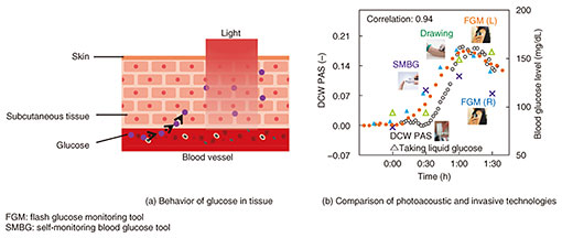
4. Application example: biological measurementsFinally, we introduce an example of applying the photoacoustic effect to the measurement of biological components. The sensor used in this example was one that sandwiches the earlobe as in the clip introduced above. We asked a healthy person to drink a beverage containing a large amount of glucose and measured subsequent change in the concentration of glucose in that person’s interstitial fluid [1, 2]. Glucose concentration in interstitial fluid differs from that in the blood (so-called blood-sugar level) in that it indicates the concentration of glucose in interstitial fluid exuded from blood vessels and supplied to cells, as shown in Fig. 4(a). It is known that the glucose concentration in interstitial fluid changes in nearly the same manner as the blood-sugar level. A comparison of measured values by using photoacoustic measurement technology with those by using various types of invasive blood-sugar-level sensors is shown in Fig. 4(b). The blue plot represents values from differential continuous-wave photoacoustic spectroscopy (“DCW PAS” in the figure), and the other plots represent values measured by drawing blood and carrying out an intravenous procedure, i.e., invasively. With the invasive technologies, the blood-sugar level increased by about 60 mg/dl 15–30 min after drinking the glucose beverage and began to decrease gradually one hour later. These results indicate that the photoacoustic method could enable measurements that conform well to this 60-mg/dl change in the blood-sugar level. By matching a target substance with an appropriate optical wavelength, this technology holds promise for application to a variety of biological components of great interest such as cholesterol, neutral fats, and other types of lipids.
5. Future workThis article introduced the potential of biological-information sensing using the photoacoustic effect and its application to technology for measuring biological components. Up to now, biochemical-information sensing has not been a target of continuous monitoring in everyday life, but if it can be made practical, we can expect it to be applied in a variety of ways. It could be used, for example, as a technology supporting online diagnostics, which have become increasingly popular during the COVID-19 pandemic. To achieve such continuous monitoring in everyday life, device size and usability will be important factors in addition to non-invasive features. Going forward, we will take up the research and development of technology that uses photoacoustic measurements to enable high accuracy analysis regardless of gas, liquid, or solid targets and that enables the continuous monitoring of biochemical information such as biological and respiratory components in everyday life. References
|
||||||